
Jennifer Curtiss, associate professor in biology at New Mexico State University, has been given a National Science Foundation award for a three-year project in morphogenesis research. The award was given to her earlier this year as a grant of $247,911.
The project is a first of its kind for Curtiss as it will be performed all on living tissue using methods and studies on live cell imaging developed by Frank Pichaud, a researcher at University College London, and a confocal microscope provided by Charles Shuster, biology professor at NMSU. The use of living tissue is crucial for Curtiss’ research as a developmental biologist.
“There's only so much we can see with dead tissue,” Curtiss said. “It's just like looking at one cartoon image instead of watching a whole movie. With living tissue, you can see the action as it's happening. We'll be able to see where a process went wrong. We can watch it in real time instead of wondering how it got from here to there.”
As a developmental biologist, she studies how single cells become whole multicellular organisms. In this process, the cells must change positions with one another in order to make the whole form. This happens through the making of the form of the organism, referred to as morphogenesis.
“Morphogenesis is the reason for things like the shape of your hand and the fact that your digits are separated from one another,” Curtiss said. “The cells themselves have to also undergo changes in their shape and the way they interact with other cells in order to make the final form. So, basically what we're trying to understand is what kinds of things do the cells have to change and how do they interact with their neighbors? What kind of forces are involved?”
After working with Pichaud and learning his methods on live cell imaging, Curtiss will return to NMSU to work with Shuster, who has years of experience with live cell imaging on confocal microscopes. The use of the confocal microscope is key to seeing morphogenesis happen in real time, and on living tissue.
Confocal microscopes use optical techniques to make optical slices of living tissue without damaging or killing the tissue. Fluorescent proteins mark certain structures under the microscope and a kind of three-dimensional model is created with the confocal microscope.
The model created can be viewed as a “movie” that shows the changes in single cells and the process of morphogenesis. This “movie” will better show Curtiss exactly what kind of forces and interactions are involved in these processes.
Curtiss recalled wanting to do a project like this for 10 to 15 years and, now that she has the chance, she’s excited to focus her research on more of an exploration of the mechanisms within the cells and how they work. This project not only benefits her and her knowledge of developmental biology, but it will also greatly benefit generations of biologists to come.
Once her work both in London and back home is complete, she plans to share these new techniques and experience with her students. She finds it critical to teach them these new methods as they will be better prepared as future researchers.
“It's super important to me,” Curtiss said. “It's so much fun to watch my students go off and do the next thing and the more that I can do for them, the better. I'm really interested in the science too, but the fun part is to work with the students on the science and watch them blossom too.”
-30-
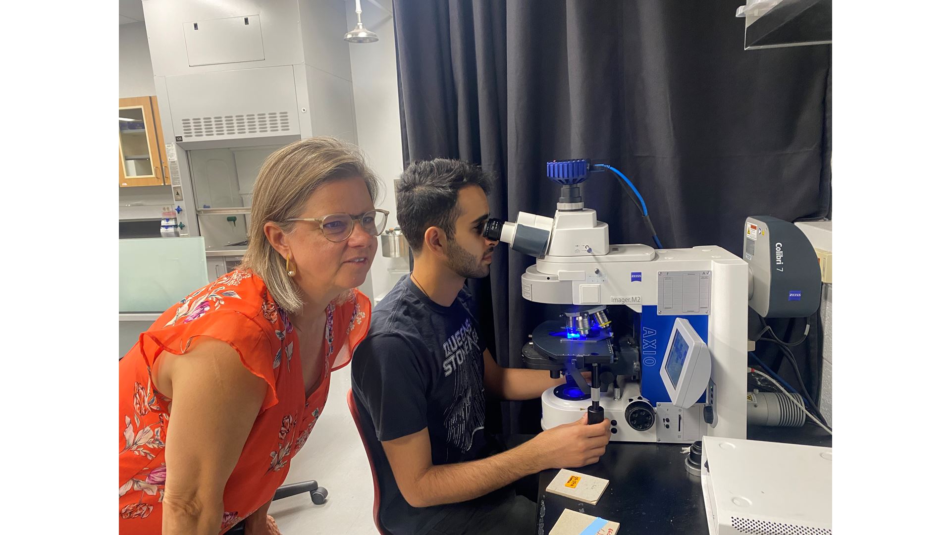
CUTLINE: Professor Jennifer Curtiss and doctoral student Abdul Al-Nouman looking at the eye of a fly under a microscope that uses different colored lights to see patterns within the cells. (NMSU photo by Chloe Dunlap)